
Fig. 2.
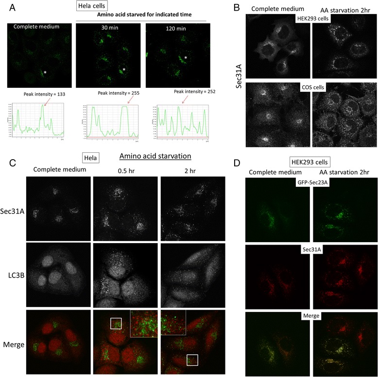
Autophagy changes the morphology of ERES. a Hela cells were cultured in growth medium or amino acid starved in EBSS to activate autophagy for indicated time. Cells were stained with ERES by anti-Sec31A antibody. Fluorescence intensity profiles of the ERES of the cells marked with asterisks were quantified. A line was drawn from a cell chosen in the image to generate fluorescence pixel intensity. b ERES in HEK293 (top) and COS cells (bottom) in complete growth medium or in EBSS (AA starvation). c Hela cells incubated with complete medium or EBSS for amino acid starvation for 0.5 or 2 h were stained with ERES (Sec31A, green in merge) and autophagosomes (LC3B, red in merge). d HEK293 cells were transfected with GFP-Sec23A (green). Transfected cells were cultured in growth medium or EBSS for 2 h before fixation and staining with Sec31A (red)