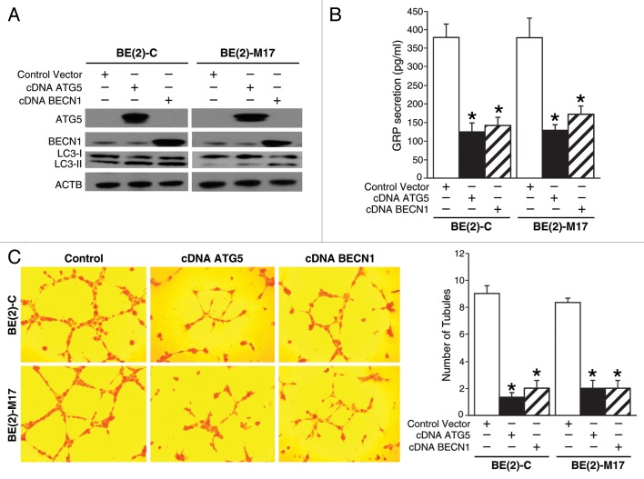
Figure 4. Overexpression of proautophagic molecules decreased GRP secretion and tubule formation by HUVECs. (A) ATG5 or BECN1 overexpression and the LC3-II, autophagosome marker, were determined by immunoblotting using lysates from BE(2)-C and BE(2)-M17 cells treated with cDNA encoding ATG5 or BECN1. ACTB was probed to assess equal loading. (B) BE(2)-C and BE(2)-M17 cells overexpressing ATG5 or BECN1 were plated on 100 mm dishes in serum-free media for 48 h, and GRP secretion was assessed by ELISA. (C) HUVECs were plated on 24-well plates coated with Matrigel and incubated with cell culture media from BE(2)-C or BE(2)-M17 cells overexpressing control vector, cDNA ATG5 or cDNA BECN1. Tubule staining was performed in triplicate and representative images shown. Values shown are mean ± SEM of three separate experiments (*P < 0.05 vs. control vector).

An official website of the United States government
Here's how you know
Official websites use .gov
A
.gov website belongs to an official
government organization in the United States.
Secure .gov websites use HTTPS
A lock (
) or https:// means you've safely
connected to the .gov website. Share sensitive
information only on official, secure websites.
