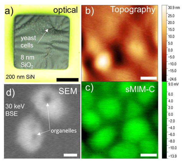
Figure 8.
(a) A wide-field optical microscopy image of yeast cells immersed in glycerol behind a 8 nm-thick SiO2 membrane. (b) sMIM topography channel shows that cell’s adhesion to SiO2 membrane affects the morphology of the membrane. (c) sMIM-C image of the same area as in (b). A larger number of visible cells is due to the probing depth of microwaves. (d) Scanning electron microscopy image of the yeast cells immersed in water solution. See details in the text. Scale bar in a is 20 μm. Scale bars in (b–d) are 1 μm.

