. 2014 Sep 16;6(1):152–157. doi: 10.1039/c4sc02380c

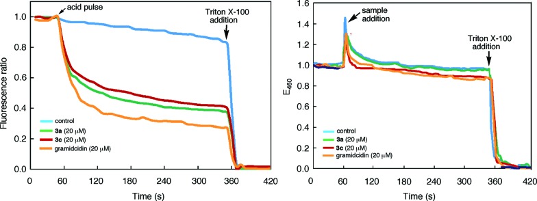
Fig. 6. (a) Time-dependent changes in the (a) ratio of the emission intensities of 8-hydroxypyrene-1,3,6-trisulfonate (HPTS, 0.1 mM) encapsulated inside large unilamellar vesicles (LUVs) of 1-palmitoyl-2-oleoyl-sn-glycero-3-phosphocholine (POPC). Solutions of LUVs (KCl 145 mM, HEPES 100 mM, pH 7.0) were first mixed with 3a, 3c, gramicidin (added from 1 mM stock solutions in THF), or THF (control) and then incubated for 2 min, followed by a HCl (2 M) pulse. The ratio of emission intensities at 510 nm by exciting at 450 nm and 405 nm respectively was monitored over 5 min; (b) the fluorescence intensity of N-(ethoxycarbonylmethyl)-6-methoxyquinolinium bromide (MQAE) encapsulated inside LUVs of POPC. Solutions of Cl free LUVs (10 mM MQAE, potassium gluconate 100 mM, HEPES 100 mM, pH 7.4) were first incubated with KCl (100 mM) in HEPES (100 mM, pH 7.4) buffer for 1 min. Stock solutions of 3a, 3c, gramicidin (1 mM in THF) and THF (control) were added to monitor the change of emission intensity at 460 nm (λ ex = 354 nm) for 5 min. The LUVs were ruptured by adding 200 μ(L of 1×X lysis buffer (1.55 mM Triton X-100 in pH 7.0 Tris-EDTA) (for (a)) or 200 μ(L of Triton X-100 (3.1 mM in H2O) (for (b)).

Fig. 6