
Figure 6.
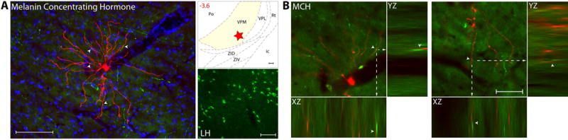
MCH innervation of thalamic trigeminovascular neurons. (A) Left: Immunopositive Melanin Concentrating Hormone axons (green) surrounding a thalamic dura-sensitive neuron (red) labeled with TMR–dextran. Nuclear counterstaining was performed with DAPI (blue). Arrowheads indicate close apposition of MCH positive axons and the cell body and dendrites of the labeled neuron. Upper right: Location of the dura-sensitive neuron (red star) shown at left. Number in red indicates distance from bregma (mm). Lower right: Fluorescent image showing MCH labeling of cell bodies in the lateral hypothalamus. Scale bars = 100 mm. (B) Close apposition between MCH immunopositive axons and thalamic trigeminovascular neurons. The three views in the x-y, y-z and x-z planes provide evidence that MCH immunopositive fibers (green) may contact distal dendrites of trigeminovascular neurons in VPM (red). Arrowheads indicate probable contact point on each view. Note that some green-labeled axons and red-labeled dendrites are in the same focal plane (yellow). Scale bar = 50 μm. Adapted from Noseda et al., 201427