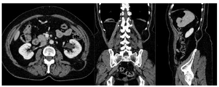
FIGURE 6. Computed tomography of abdomen revealed protrusion of retroperitoenal fat through the muscles in the upper lumbar triangle topography, defect of about 18 mm (major axis); hernial sac of about 54 mm which was located between the lateral border of the erector muscle of the spine and the medial margin of the internal oblique muscle under the latissimus dorsi muscle, characteristic finding ofGrynfelthernia.

