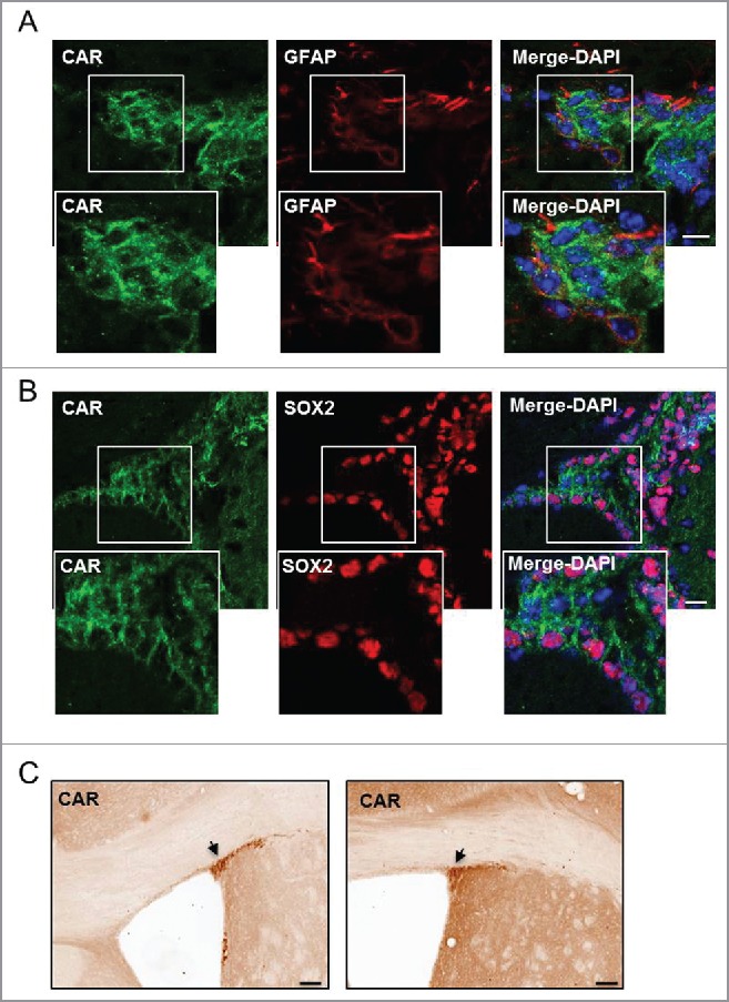
Figure 1.

CAR in the SVZ. (A and B) Healthy mouse brains were stained with anti-CAR, anti-GFAP or anti-SOX2 and DAPI. (A) CAR (in green) was found in few radial glial cells of the SVZ identified by GFAP (in red) (white box refers to zoomed in panels). (B) The majority of CAR+ cells in the SVZ express SOX2 (in red), which correspond to proliferating NPCs (white box refers to zoomed in panels). (C) Representative image of anti-CAR immunohistochemistry in the murine SVZ. Black arrows show areas of intense CAR staining in the SVZ. Scale Bars (A and B) 20 μm (C) 200 μm.
