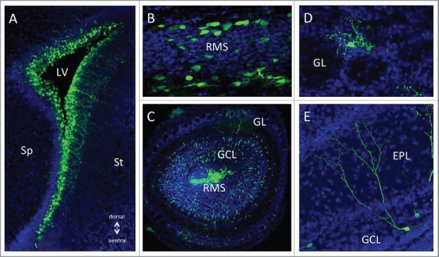
Figure 2.

GFP expression from CAV-GFP infected cells in the postnatal forebrain neurogenic system. 1 × 10⁹ physical particles of CAVGFP33 was injected in the lateral ventricle (LV) of P1 mice. (A) 1 day post-injection (dpi) exclusively radial glia type neural stem cells located in the ventricular zone surrounding the lateral ventricle (LV) expressed GFP. Over the following days, tangentially migrating neuroblasts in the rostral migratory stream (B) and radially migrating neuroblasts in the olfactory bulb (OB, C) were GFP+. After arrival in the olfactory bulb (at 20 dpi) newly generated neurons in the glomerular layer (GL, D) and the granule cell layer (GCL, E) showed strong and long-lasting GFP expression. EPL; External Plexiform Layer, Sp; septum, St; striatum.
