Figure 3.
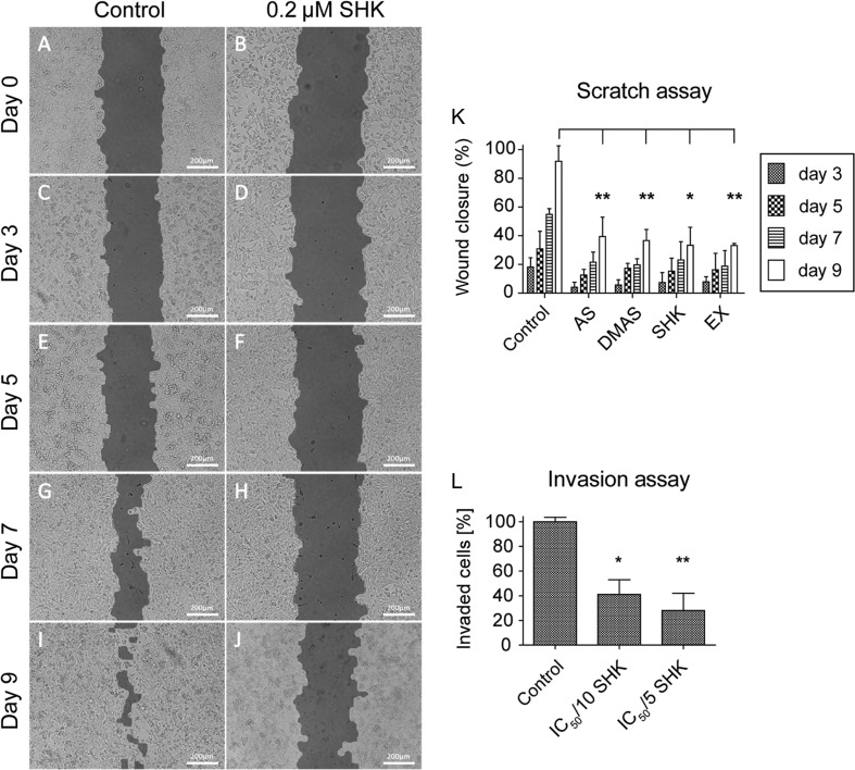
Shikonin derivatives inhibit the migration and invasion of TT cells (A, B, C, D, E, F, G, H, I and J). In a monolayer wound-healing assay, solvent control-treated TT cells were able to close the gap (images on the left), whereas TT cells treated with the corresponding IC50/5 concentration of each compound failed to do so. Representative images show TT cells treated with DMSO (control) and the IC50/5, i.e. 0.2 µM shikonin (SHK, images on the right). Images were acquired immediately after the scratch was created (A and B), 3 days (C and D), 5 days (E and F), 7 days (G and H) and 9 days (I and J) later. Scale bars, 200 µm. (K) Statistical analysis of the scratch assay. All tested compounds showed similar efficiency on the inhibition of wound closure. (L) Treatment with 0.1 µM (IC50/10) shikonin reduced the invasion capacity of TT cells. AS, acetylshikonin; DMAS, β,β-dimethylacrylshikonin; SHK, shikonin; EX, O. paniculata extract; *P < 0.05, **P < 0.01, n = 3.

 This work is licensed under a