
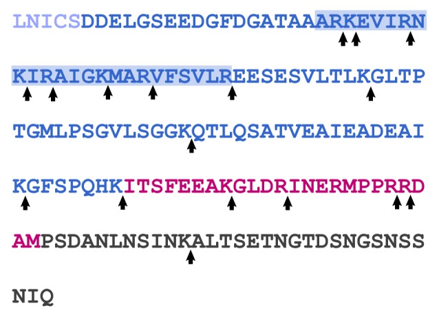
Figure 3. Sequence of the RD (dark blue), AID (red), and CT (dark gray) from human CnA. The CaM binding region is denoted by the blue-shaded box. Potential trypsin cleavage sites are noted by arrows.
Official websites use .gov
A
.gov website belongs to an official
government organization in the United States.
Secure .gov websites use HTTPS
A lock (
) or https:// means you've safely
connected to the .gov website. Share sensitive
information only on official, secure websites.
Figure 3. Sequence of the RD (dark blue), AID (red), and CT (dark gray) from human CnA. The CaM binding region is denoted by the blue-shaded box. Potential trypsin cleavage sites are noted by arrows.