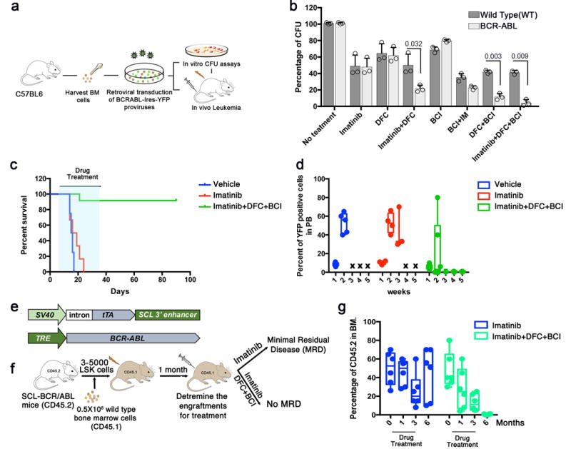
Figure 3. Chemical inhibition of c-Fos, Dusp1 and BCR-ABL eradicates minimal MRD in mice. a.

Experimental design for testing the efficacy of small molecule inhibitors of c-Fos (difluorocurumin; DFC) and Dusp 1 ((E)-2-Benzylidene-3-(cyclohexylamino)-2,3-dihydro-1H-inden-1-one; BCI) in vitro and in vivo in CML. b. Percent CFUs from WT and BCR-ABL LSK (Lin−Sca1+Kit+) cells treated with the indicated drugs. The data shown are the mean colony number from two independent experiments ± S.D. (n=3, P values are indicated above the compared bars). c. Survival curve of BCR-ABL-expressing Kit+ cell recipients treated with vehicle (blue), imatinib (red), or a combination of imatinib with DFC and BCI (green). The time period during which the drugs were administered is indicated by light blue shading. Data shown are from one of the two independent transplantation experiments with similar results (n = 5 mice per group; p = 0.0285). d. Percent of YFP+ cells in peripheral blood of mice treated with imatinib or Imatinib+DFC+BCI. e. Schematic structures of the transgenes used in transgenic mice to drive BCR-ABL expression in stem cells. Top, the Scl-3′enhancer drives expression of the tetracycline transactivator-protein (tTA); bottom a tetracycline-responsive promoter (Tet-P) drives BCR-ABL expression. Transgenic mice are fed doxycycline-containing chow; doxycycline withdrawal induces expression of BCR-ABL in hematopoietic stem cells, generating leukemic stem/progenitor cells. f. Experimental design for studying the effects of Dusp1 and c-Fos inhibition in leukemic stem cells. Mice received a competitive transplant of 3,000–5,000 LSK Scl-BCR-ABL cells with 500,000 wild type (WT) total bone marrow cells. Engraftment was evaluated one month after transplantation by flow cytometry (CD45.1 vs. CD45.2); the mice were then treated with imatinib alone or imatinib+DFC+BCI for three months, and the presence of MRD was evaluated at indicated times. g. Percentage of leukemic cells (CD45.2) in bone marrow of BoyJ recipients (CD45.1) at the indicated time points after cell transplantation. Imatinib treatment (blue bars) reduces leukemic burden but not curative treatment with imatinib+DFC+BCI completely eradicated the leukemic cells (green bars). Individual data points are denoted as empty circles in each bar graphs.
