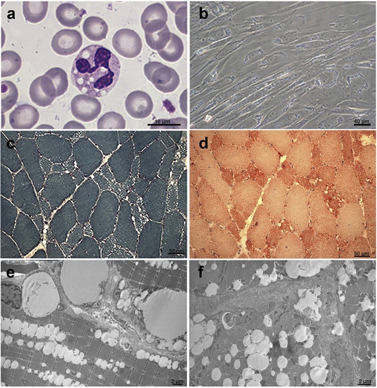
Fig. 2.
Histochemical characterization of NLSDM patient. (a) Detection of Jordans' bodies in granulocytes, stained with May-Grünland Giemsa (MGG). (b) Phase contrast image of cultured fibroblasts from the patient reveals increase of lipid droplet storage inside the cells. Consecutive cryosections of the patient muscle biopsy, stained with Gomori trichrome (c) and Oil-Red-O (d), show microvacuoles and abnormal accumulation of lipids. (e,f) Electron microscopy reveals massive line-up of lipid droplets without signs of mitochondrial alteration.

