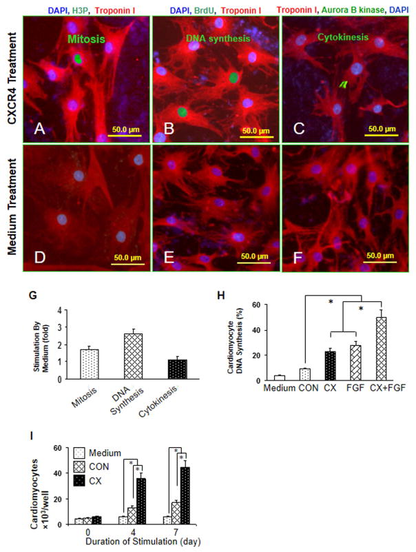
Fig. 1. MSCCX4 conditioned medium induces proliferation of neonatal cardiomyocytes.
Primary neonatal rat ventricular cardiomyocytes were treated with concentrated media from MSCCX4. Cell cycle activity was detected with an antibody directed against phosphorylated histone H3 (H3P) for mitosis (A, D), BrdU for DNA synthesis (B, E), and aurora B kinase for cytokinesis (C, F) and were quantified by immunofluorescence microscopy (G). The additive effect of media from MSCCX4 plus FGF (100ng/ml) on DNA synthesis was observed (H). Neonatal cardiomyocyte proliferation was determined by cell counting (I). Color codes are given at the top. Data are means ± SEM of 20 independent experiments. *p<0.05; CX indicates medium from MSCCX4 group; CON, medium from MSCNull group.

