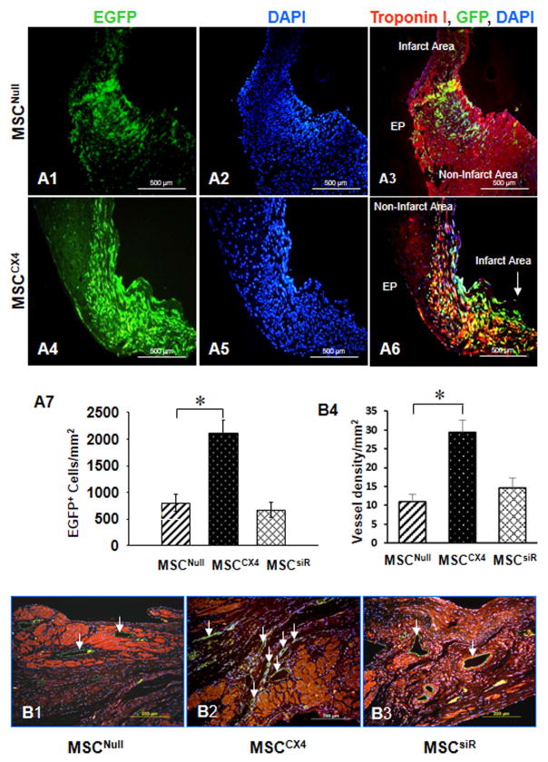
Fig. 5. Effects of MSCCX4 transplantation in the infarct area at 4 weeks post MI.
(A) Migration of the EGFP positive cells was observed in MSCNull group (A1–A3) and in MSCCX4 (A4–A6). Color codes given at the top. Original magnification: ×100. EGFP migration was estimated quantitatively in infarcted myocardium from various treatment groups (A7). (B) MSCCX4 group promotes vessel formation by significant expression of CD31 (green color (white arrow) (B2) in comparison with MSCNull (B1) or MSCsiR group (B3), (magnification×200). Quantitative analysis of capillary density (anti-CD31 antibody staining) was determined by various treatments (B4). Data are means ± SEM of 6 independent experiments. *p<0.05 vs. MSCNull.

