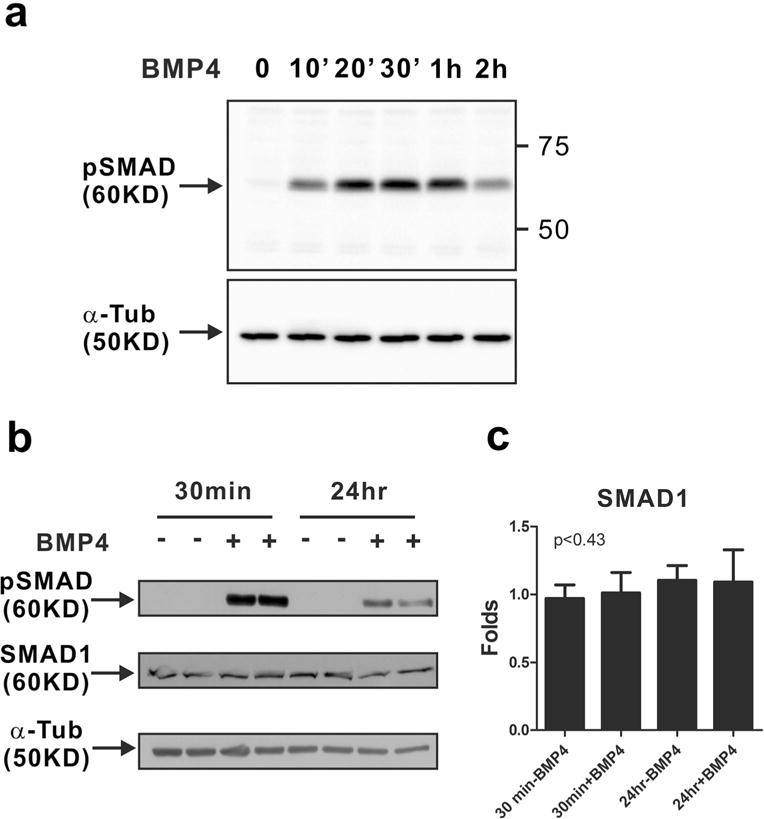
Fig. 1.

Activation of canonical BMP signaling in MCF-10A cells in response to BMP4. MCF-10A cells were treated with BMP4 (50 ng/ml) after serum starvation. Western blot analysis of phosphorylated SMAD1/5/9 (pSMAD) was performed. a The phosphorylation of SMAD1/5/9 (pSMAD) was detectable 10 min after BMP4 treatment. Alpha-tubulin (α-Tub) was used as an internal control in Western blots of phosphorylated SMAD1/5/9. b The phosphorylation of SMAD1/5/9 was observed 24 h after BMP4 treatment as well as 30 min. Western blot analysis of phosphorylated SMAD1/5/9 (pSMAD) and SMAD1 was performed. α-Tub was used as an internal control in the western blot. c The band intensity of SMAD1 was quantified by ImageJ and normalized to α-tubulin levels. The graph shows fold increase as compared with the levels in the non-treated control group (30 min without BMP4 treatment). Results are shown in mean±SD, and p value was calculated with one-way ANOVA
