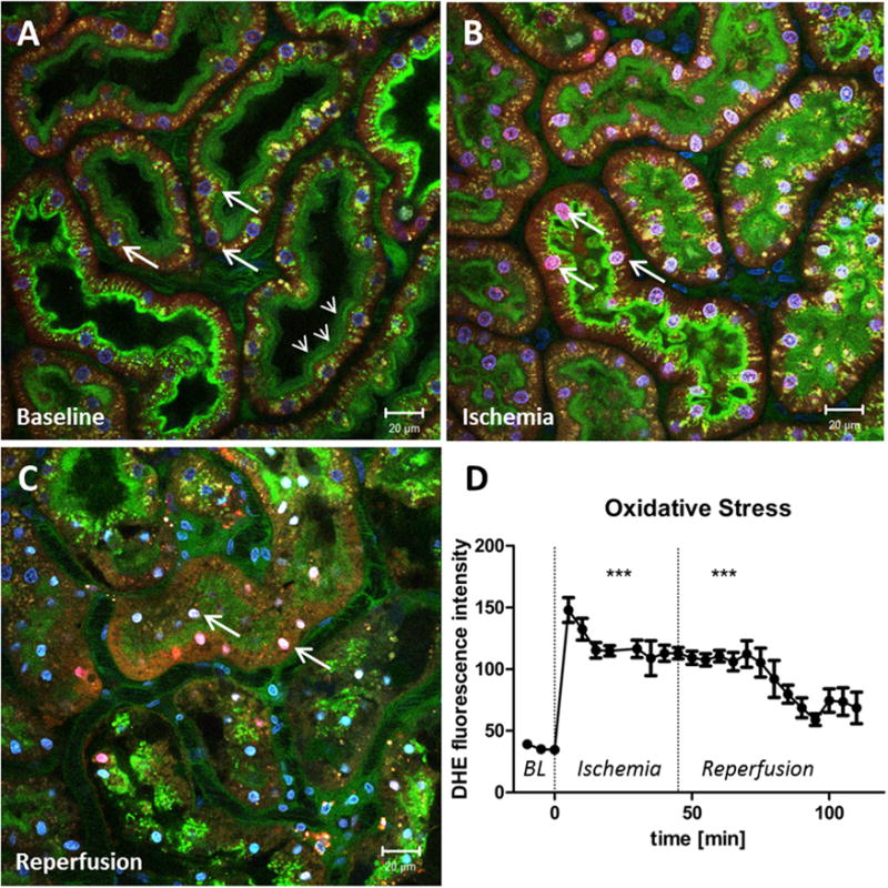
Figure 2.

Oxidative stress in proximal tubules is enhanced by ischemia. (A) Renal cortex of a MWF rat during baseline conditions. The peritubular vasculature is labeled green by the injection of fluorescein isothiocyanate albumin. Glomerular-filtered fluorescein isothiocyanate albumin is reabsorbed in the PTs and stains the brush border green (small arrows). Cell nuclei are stained blue by Hoechst 33342 (big arrows). To indicate the generation of reactive oxygen species, DHE was injected into the jugular vein, which is activated to a red dye by superoxide and then translocates to the nucleus. Before ischemia the DHE staining of the PT nuclei is weak and oxidative stress is low. However, DHE intensity of the PT nuclei rapidly increases at the onset of ischemia (arrows in panel B) and remains increased during reperfusion (arrows in panel C). (D) Nuclear fluorescence intensity of DHE in PTs as a function of time before, during, and after ischemia. ***DHE fluorescence is significantly higher during ischemia and reperfusion, when compared with baseline (BL) values (P > .0001).
