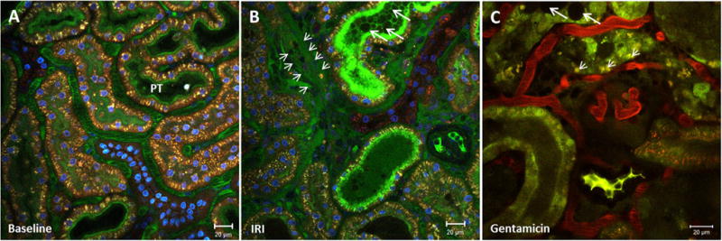
Figure 3.

Visualization of apical membrane blebbing and cell shedding. Renal cortex of a MWF rat (A) before and (B) after a 45-minute ischemia period. The peritubular vasculature is stained green by fluorescein isothiocyanate albumin and the nuclei blue by Hoechst 33342. (B) IRI induces necrotic cell death in proximal tubules and leads to apical membrane blebbing, cell shedding, and the release of DAMPs, which accumulate in the tubular lumen and form tubular casts (big arrows). In areas of diminished peritubular blood flow, extravasation and edema formation is visible by the appearance of fluorescein isothiocyanate albumin in the interstitium (small arrows). (C) Renal cortex of a gentamicin-treated rat (100 mg/kg on 5 consecutive days). Proximal tubular dysfunction can be detected by enlarged lysosomes within the PT cells (small arrows). Apical membrane blebbing, shedded cells, and DAMPs form tubular casts (big arrows) in the lumen of some PTs.
