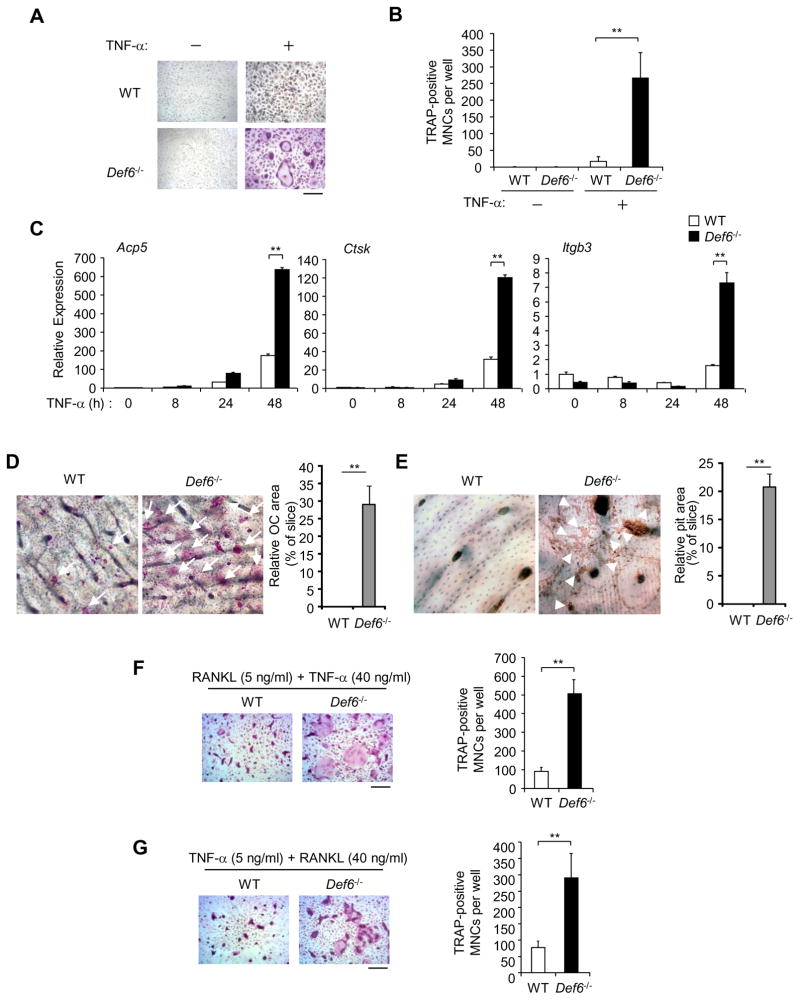
Figure 3. Def6 deficiency increases TNF-α-induced osteoclast formation.
(A) BMMs derived from WT control and Def6−/− mice were treated with TNF-α (40 ng/ml) for 3 days. TRAP staining was performed (A) and the number of TRAP-positive MNCs per well was counted (B). TRAP-positive cells appear red in the photographs. Scale bar, 100μm. Data are representative of at least five independent experiments. **P<0.01. (C) Quantitative real-time PCR analysis of mRNA expression of Acp5 (encoding TRAP), Ctsk (encoding cathepsin K) and Itgb3 (encoding β3) in BMMs from the WT control and Def6−/− mice treated with TNF-α (40 ng/ml) for the indicated times. Data are representative of at least three independent experiments. **P<0.01. (D) TRAP staining of bone slices (left panel) on which the WT control and Def6−/− BMMs were cultured with TNF-α for 5 days. Osteoclasts formed on the bone slices appeared red by TRAP-staining, and were also indicated by white arrows. Right panel: the relative area of the osteoclasts (OCs) to the slice (%). **P<0.01. (E) Bone resorption indicated by yellow-red stained pits on the bovine bone slices by wheat germ agglutinin staining, which was also indicated by white arrowheads. Right panel: the relative area of the pits to the slice (%). **P<0.01. (F) The WT control and Def6−/− BMMs were treated with RANKL (5 ng/ml) for 1 day followed by co-stimulation with RANKL (5 ng/ml) and TNF-α (40 ng/ml) for 2 days. (G) The WT control and Def6−/− BMMs were treated with TNF-α (5 ng/ml) for 1 day followed by co-stimulation with TNF-α (5 ng/ml) and RANKL (40 ng/ml) for 2 days. Osteoclast formation was determined by TRAP staining (left panels in F and G) and the number of TRAP-positive MNCs per well (right panels in F and G). **P<0.01.

