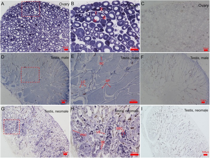
Fig 4. Cyto-locations of CS-β-catenin1 mRNA in gonads of C. semilaevis.
(A). Low magnification showing the adult ovaries. (B). High magnification of the framed area in (A). (C). Control of β-catenin1 localization in female ovaries. (D). Low magnification showing the male adult testis of males. (E). High magnification of the framed area in (D). (F). Control of β-catenin1 localization in male testis. (G). Low magnification showing the adult testis of a neomale. (H). High magnification of the framed area in (G). (I). Control of β-catenin1 localization in neomale testis. Three samples were handled. Oocytes at different developmental stages are marked by I, II, III and IV. PSC: primary spermatocytes; SSC: secondary spermatocytes; LE: Leydig cells; SP: spermatid; SE: Sertoli cells. Scale bars: 100 μm.

