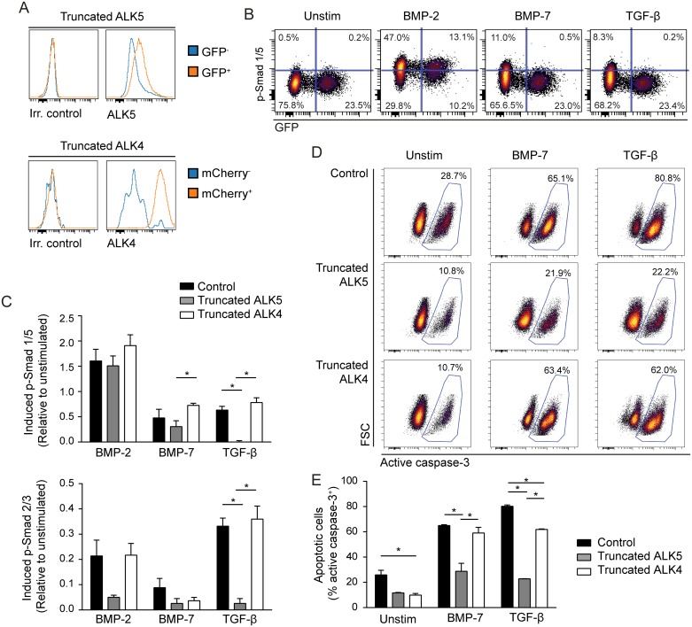
Fig 5. Introduction of truncated ALK5 in the B-cell lymphoma cell line Mino abrogates BMP-7-induced apoptosis.
Mino cells were transduced with truncated ALK5 or truncated ALK4. (A) The cells were stained with biotinylated anti-ALK5, followed by streptavidin PE or by biotinylated anti-ALK4, followed by streptavidin APC, and analyzed by flow cytometry. Receptor expression is compared in GFP+ or mCherry+ transduced cells vs. GFP-/mCherry- non-transduced cells. (B-C) The transduced Mino cells were cultured in serum free media (X-VIVO 15) over night and then left in medium alone (unstim) or stimulated with BMP-2, BMP-7 or TGF-β for 60 min, before detection of phosphorylated (p-) Smad 1/5 or p-Smad 2/3 by flow cytometry. (B) One representative experiment showing p-SMAD1/5 vs. GFP in truncated ALK5-2A-GFP expressing cells. (C) BMP- or TGF-β-induced phosphorylation is shown relative to unstimulated cells, using arcsinh transformation of median fluorescence intensity data. Mean ± SEM, n = 5. (D-E): Transduced Mino cells were cultured in X-VIVO 15 and left unstimulated or stimulated with TGF-β or BMP-7 for 72 hours and stained for active caspase-3 before analysis by flow cytometry. Shown here is active caspase-3 staining of control cells and transduced cells for (D) one representative experiment and (E) mean ± SEM, n = 3. *p < 0.05; two-tailed, paired Student’s t-test.

