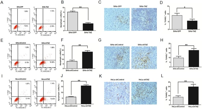
Fig 5. TAZ inhibits apoptosis in cervical cancer cells both in vitro and in vivo.
Apoptosis of TAZ-mediated cervical cancer cells was monitored with fluorescence-activated cell sorting (FACS) analysis. The representative cell apoptosis histograms and the percentage of apoptosis cells of TAZ-mediated cervical cancer cells are shown: (A, B) SiHa-GFP and SiHa-TAZ cells; (E, F) SiHa-shControl and SiHa-shTAZ cells; (I, J) HeLa-shControl and HeLa-shTAZ cells. Values are expressed as the mean±SD of three experiments in duplicate. Apoptotic cell death in tumor xenografts formed by TAZ-mediated cervical cancer cells was measured by TUNEL assay, and representative micrographs are shown (magnification, ×40):(C) SiHa-GFP and SiHa-TAZ cells; (G) SiHa-shControl and SiHa-shTAZ cells; (K) HeLa-shControl and HeLa-shTAZ cells. Quantitative analysis of apoptosis cells in xenograft samples formed by TAZ-mediated cervical cancer cells: (D) SiHa-GFP and SiHa-TAZ cells; (H) SiHa-shControl and SiHa-shTAZ cells; (L) HeLa-shControl and HeLa-shTAZ cells. Six tumor samples were measured and analysed every group. Values are shown as the mean±SD. *P<0.05 vs. control; ** P<0.01 vs. control.

