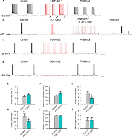
Fig. 5. The effect of the ERG activator on burst discharges of subthalamic neurons recorded at a membrane potential of about −65 mV.

The horizontal dashed lines indicate the level of −65 mV. (A) A representative neuron that spontaneously fires in bursts with relatively long plateau is shown. PD-118057 (0.5 μM) evidently hyperpolarizes the burst plateau and also shortens burst duration [see analyses in (E) to (J), n = 4; see also figs. S4B and S6B]. (B to D) Three representative neurons that spontaneously fire in relatively short bursts are shown [n = 8, 8, and 4 for (B), (C), and (D), respectively]. Note that the baseline membrane potential is not markedly altered by PD-118057 (0.5 μM) in any case but is slightly depolarized by E-4031 (5 μM). (B) The burst pattern remains in 0.5 μM PD-118057 but is abolished by the addition of 5 μM E-4031. (C and D) PD-118057 (0.5 μM) hyperpolarizes the plateau and abolishes bursts. (E to J) In subthalamic neurons having burst duration >3 s, there are no significant effects of PD-118057 (0.5 μM) on burst rates (E), intraburst spike frequency (F), the lowest potential (I), and AHP amplitude (J). However, PD-118057 significantly shortens the burst duration (G; see also fig. S9B) and decreases the number of spikes per burst (H) (n = 4; *P < 0.05; **P < 0.01 compared to control, paired two-tailed Student’s t test). Scale bars, 20 mV/2 s. Animals used: p18 to p26 mice.
