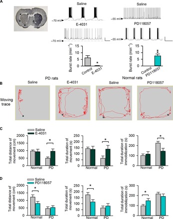
Fig. 6. Remedy of locomotor deficits of parkinsonian rats by ERG channel blockers.

(A) Left: Tyrosine hydroxylase immunohistochemistry shows unilateral dopaminergic deficiency (indicated by arrow heads) in the striatum of parkinsonian (PD) rat coronal brain sections. Middle and right: Whole-cell recording in STN slices from either PD rats [where subthalamic neurons tend to fire in the burst mode (middle); refer to more cases in fig. S4] or normal rats [where subthalamic neurons more frequently fire in the tonic mode (right)] in the absence and presence of 5 μM E-4031 (n = 5; middle) and 0.5 μM PD-118057 (n = 7; right), respectively. Scale bars, 20 mV/2 s. *P < 0.05; **P < 0.01, paired two-tailed Student’s t test. Also refer to in vivo electrophysiological data in fig. S11. (B) Representative locomotor traces of a PD (left) or a normal (right) rat in an arena with administration of normal saline and 200 μM E-4031 (left) or PD-118057 (right), respectively. (C and D) Changes in locomotor activities after direct microinjection of E-4031 or PD-118057 into the STN. The changes in the total distance of movement (left), total duration of movement (middle), and total duration of immobilization (right) in normal or parkinsonian (PD) rats with administration of either 200 μM E-4031 (C; n = 10 for both normal and PD groups) or 200 μM PD-118057 (D; n = 9 for both normal and PD groups) are compared with those injected with normal saline. *P < 0.05, two-way mixed-design analysis of variance (ANOVA). Application of E-4031 markedly improves locomotor activities of PD (but not normal) rats in (C). In contrast, PD-118057 significantly decreases locomotor activities of normal (but not PD) rats in (D). See also fig. S10. Animals used: p37 to p40 rats for brain slice recording; 2- to 3-month-old rats (weighing 260 to 380 g) for other experiments.
