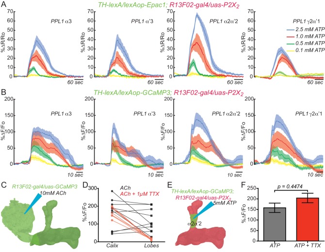
Figure 2. KC and DAn PPL1 neurons axoaxonic reciprocal synapses are functional.
(A) Cyclic AMP responses in DAn (α3, α’3, α2α’2 and γ2α’1) to artificial stimulation of KC using ATP/P2X2 system. The solid line of each trace represents the mean and shaded area represents ± SEM at various concentrations of ATP used in the experiment. Cyclic AMP accumulation was observed in the axons of the DAn that innervate all of the MB lobe compartments that were recorded. N = 9–14. (B) Calcium responses in DAn (α3, α’3, α2α’2 and γ2α’1) to artificial stimulation of KC using ATP/P2X2 system. The solid line of each trace represents mean and shaded area represents ± SEM. Calcium responses were observed in the axons of the DAn that innervate all of the MB lobe compartments that were recorded. N = 12–16. (C) Diagram of the experimental setup. A micropipette was used to focally apply ACh to the calyx of the MB while imaging in both the calyx and MB lobes. (D) Calcium responses in both calyx and lobes of the KC to ACh application in the presence (red) or absence (black) of 1 µM TTX. TTX blocked the responses in the lobes but not the calyx, indicating that TTX was functional in blocking action potentials. N = 7–10. (E) Diagram of the experimental setup. A micropipette was used to focally apply 5 mM ATP to the α2α2’ compartment of MB while recording calcium responses in α2α’2 DAn. (F) Calcium responses in α2α’2 DAn in the presence (red) or absence (gray) of 1 µM TTX. TTX was without significant effect, indicating that the artificial activation of KC fibers were capable of evoking responses in DAn axon terminals through local, mono-synaptic transmission. N = 9–11. Data were analyzed using Mann-Whitney U non-parametric test. Bars represent the means ± SEM.


