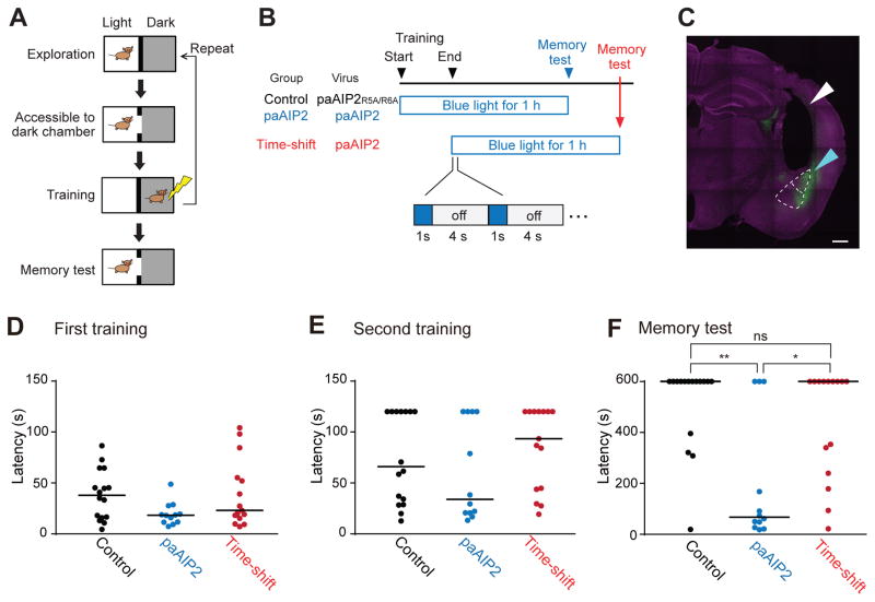
Fig. 7. Inhibition of CaMKII by paAIP2 impairs memory formation in inhibitory avoidance learning.
A. A schematic of double-trial training inhibitory avoidance. Upon entering and exploring the light chamber within the inhibitory avoidance apparatus, a mouse is allowed to enter the dark chamber, after which a foot shock is administered. The training is repeated once more. Each training trial ends if the animal crosses or avoids the dark chamber for 120 s.
B. A schematic of light application during inhibitory avoidance. For the control and paAIP2 groups, blue light (0.2 Hz, 1s) is applied from the onset of training for an hour. For the time-shift group, light delivery begins after the training. Timelines are not drawn to scale.
C. The expression of mEGFP-P2A-paAIP2 in amygdala. The cyan and white triangles indicate the area of transgene expression and the fiber track above amygdala, respectively. The upper region demarcated by white dotted line indicates lateral amygdala (LA). The lower region indicates basolateral amygdala (BLA). Scale bar = 500 μm. Note that animals expressing detectable transgene expression either in LA or BLA in both hemispheres were included in the analyses D–F.
D, E. Cross latency for the first (A) and second (B) training trials. Three groups of mice show no statistically significant difference in cross latency in both training trials. Cutoff latency set at 120 s. Bars represent median. Dunn’s multiple comparisons test following Kruskal-Wallis test (n = 16, 12 and 15 for control, paAIP2, and time-shift group, respectively).
F. Cross latency for the memory test (1 h). Cutoff latency set at 600 s. Bars represent median. P-values are from Dunn’s multiple comparisons test following Kruskal-Wallis test.

