
Fig. 2.
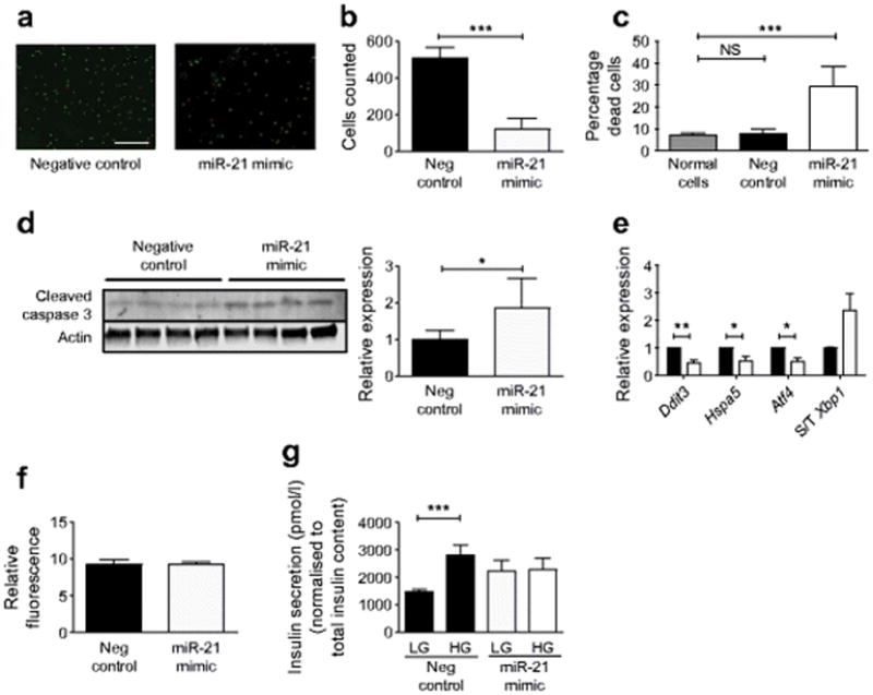
miR-21 overexpression increases INS-1 beta cell apoptosis and decreases GSIS. INS-1 cells were transfected with a miR-21 mimic for 48 h to increase miR-21 activity. (a) Viability staining with AOPI, with green staining (AO) representing all cells, and red staining (PI) representing compromised membranes. Scale bar, 400 μm. (b) Total cells and (c) percentage of dead cells were analysed. (d) Immunoblot for cleaved caspase-3. (e) Relative expression of ER stress transcripts was quantified with qPCR: black bars, control cells; white bars, mimic. (f) ROS generation was quantified using CellROX Deep Red Fluorescent Reagent. (g) After miR-21 mimic transfection, GSIS was assayed in response to low (2.5 mmol/l) and high (15 mmol/l) glucose concentrations, and normalised to the total insulin content of the cell lysate. Neg, negative. S/T Xbp1, spliced/total Xbp1. n=4–9. *p≤0.05; **p≤0.01, ***p≤0.001