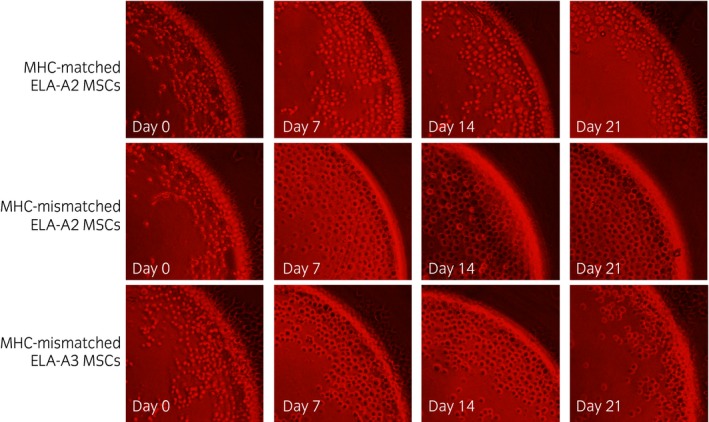
Figure 2.

10× images from Terasaki plate wells used for microcytotoxicity assays containing equine leucocyte antigen (ELA)‐A2 mesenchymal stem cells (MSCs) or ELA‐A3 MSCs and neat antisera collected on Days 0, 7, 14, or 21 post‐injection with either major histocompatibility complex (MHC)‐matched or MHC‐mismatched MSCs. Live cells appear round with a clear centre. Dead cells appear flat with a dark centre. Cell death was estimated to be <10% for MHC‐matched wells on all days and for MHC‐mismatched wells on Day 0 as shown in this figure. Cell death was estimated to be >80% for all MHC‐mismatched wells on Days 7–21 as shown in this figure.
