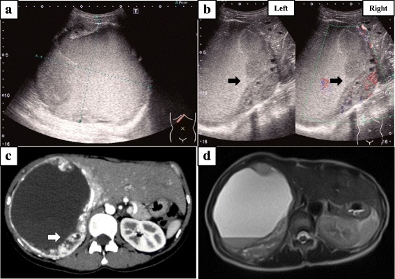
Fig. 1.

Findings from abdominal ultrasonography, CT, and MRI. a Abdominal ultrasonography shows an extensive space-occupying lesion in the right lobe of the liver, 15 cm in diameter. b Left photo shows a mural nodule, and right photo shows a heterogeneous internal component including hemorrhage and hypervascularity (black arrow). c Abdominal contrast-enhanced computed tomography shows a cystic mass measuring 13 × 14 × 11 cm in the right lobe of the liver with an enhanced mural nodule (white arrow). d Abdominal magnetic resonance imaging (MRI) shows a hyperintense component on T2-weighted imaging compatible with the hemorrhagic area
