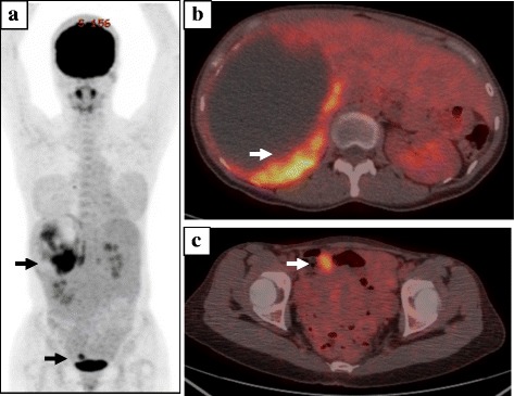
Fig. 2.

Findings of FDG-PET. a The fusion image of FDG-PET shows abnormal accumulation in the liver and lower abdomen (black arrows). b Cross-section of the upper abdomen indicates abnormal accumulation in the mural nodule in the liver (white arrow). c Cross-section of the lower abdomen indicates abnormal accumulation of the omental tumor (white arrow)
