Fig. 4.

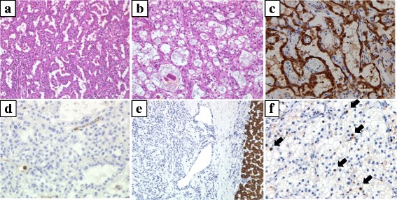
Histological findings of tumors. a Histological specimen of liver tumor (hematoxylin and eosin (HE), ×200) shows epithelioid-type mesothelioma cells with tubular components. b Mesothelioma cells with cystic components (HE, ×200). c Immunohistochemical staining for calretinin shows positive tumor cells (×200). d Immunohistochemical staining for CEA shows negative tumor cells (×200). e Immunohistochemical staining for HepPer1 shows negative tumor cells (×200). f Immunohistochemical staining for Ki-67, a marker of tumor proliferation, shows positive tumor cells (brown nuclei indicated with black arrows). Ki-67 index is 5–6%
