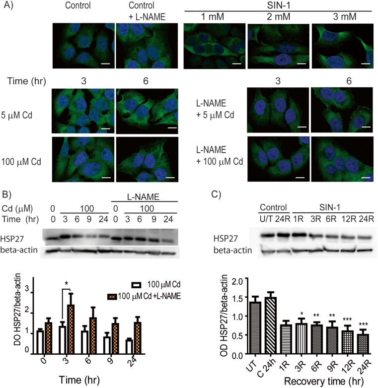
Fig. 2.
SIN-1 and Cd lowered the levels of HSP27 in HeLa cells. a Nitrotyrosine images were obtained by confocal microscopy. Top panel: Representative images of untreated HeLa cells (basal control), cells pre-treated with L-NAME (500 μM; similar to untreated), and cells treated with SIN-1 (1–3 mM) for 3 h plus 24 h recovery (positive control) are shown. Bottom panel: Nitrotyrosine was evaluated in HeLa cells exposed to Cd (5 or 100 μM) ± L-NAME pre-treatment. Green: nitrotyrosine; blue: DAPI; bar: 8 nm. b Western blots showing HSP27 expression in HeLa cells exposed to 100 μM Cd (3–24 h) + pre-treatment with L-NAME. c Western blots of HeLa cells exposed to 1 mM SIN-1 for 3 h and then assayed for HSP27 expression after 1–24 h of recovery. c, d Densitometries of the HSP27/beta-actin bands were measured and graph values represent the means of three independent experiments ± SD (*p < 0.05; **p < 0.01; ***p < 0.001) (color figure online)

