
Figure 1.
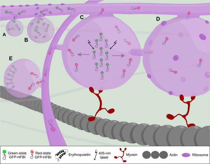
A working model of protein body formation and development. (A) Proteins are synthesized on the rough endoplasmic reticulum (ER) by ribosomes and transferred into the ER lumen co‐translationally. Protein body formation initiates when localized high concentrations of recombinant proteins occur in the ER lumen. High concentration of proteins is represented by several GFP‐HFBI molecules in an area (GFP‐HFBI is presented here as an example. These properties can be generalized to other protein fusions as well). (B) Once protein bodies (PBs) form, they grow in size over time and store higher amounts of proteins in their lumen. Co‐expression of high‐value recombinant proteins, in this case erythropoietin (EPO), with GFP‐HFBI results in passive sequestration of EPO molecules into GFP‐HFBI‐induced PBs. (C). PBs remain connected with the ER and exchange their content with other PBs via the ER or by direct contact. (D) PBs are part of the rough ER and studded with ribosomes. These ribosomes may contribute to accumulation of proteins in PBs. (E) The ER network is essential in connecting PBs that are located far from each other. In this model, GFP‐HFBI photoconverts to red‐state GFP‐HFBI upon irradiation (shown with arrows) (C). Photoconverted proteins move from one PB to neighbouring PBs (D) and to far away PBs through the ER (E). The ER and PB movement relies on the actomyosin cytoskeleton.