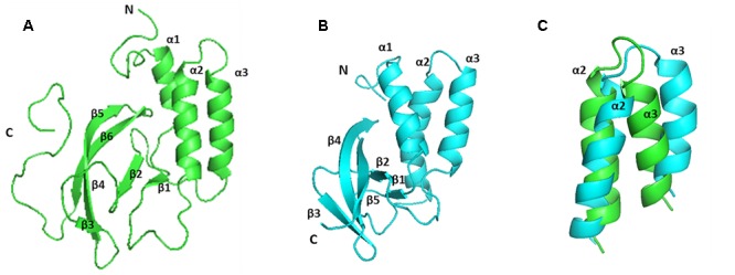
FIGURE 5.

Comparison of the solution structure of PRRSV and EAV NSP7α proteins. (A) The structure of PRRSV NSP7α (PDB accession number: 5I65). (B) The structure of EAV NSP7α (PDB accession number: 2L8K). (C) Overlay of the region from helices α2 to α3. PRRSV NSP7α is colored in green and EAV NSP7α is in blue.
