FIGURE 1.

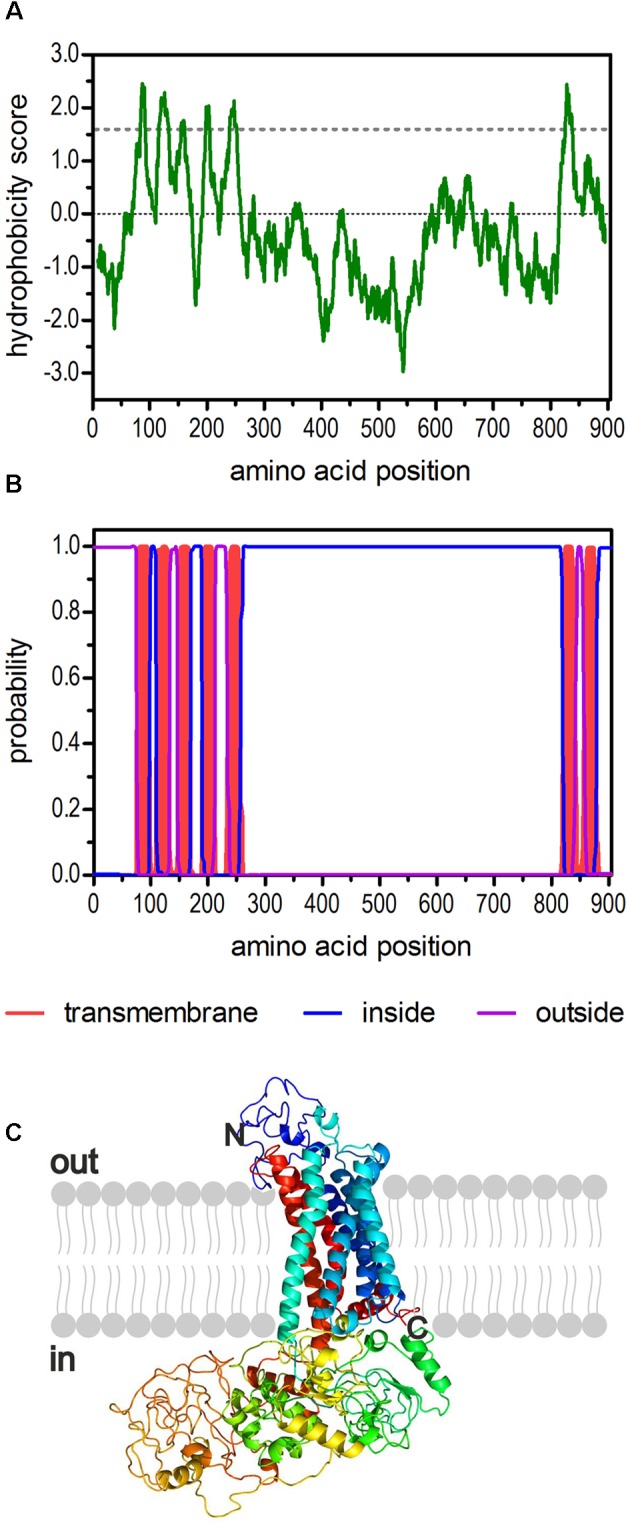
Structural characteristics of the deduced amino acid sequence of Dm5-HT2B. (A) Hydrophobicity profile of Dm5-HT2B. The profile was calculated according to the algorithm of Kyte and Doolittle (1982) using a window size of 19 amino acids. Peaks with scores greater than 1.6 (dashed line) indicate possible transmembrane regions. (B) Prediction of transmembrane domains with TMHMM server v. 2.0 (Krogh et al., 2001). Putative transmembrane domains are indicated in red. Extracellular regions are shown as purple line, intracellular regions as blue line. (C) The primary sequence of Dm5-HT2B was submitted to Phyre2 (Kelley et al., 2015). The 3D model of the receptor is color-coded (rainbow). The extracellular N-terminus and the intracellular C-terminus are labeled.
