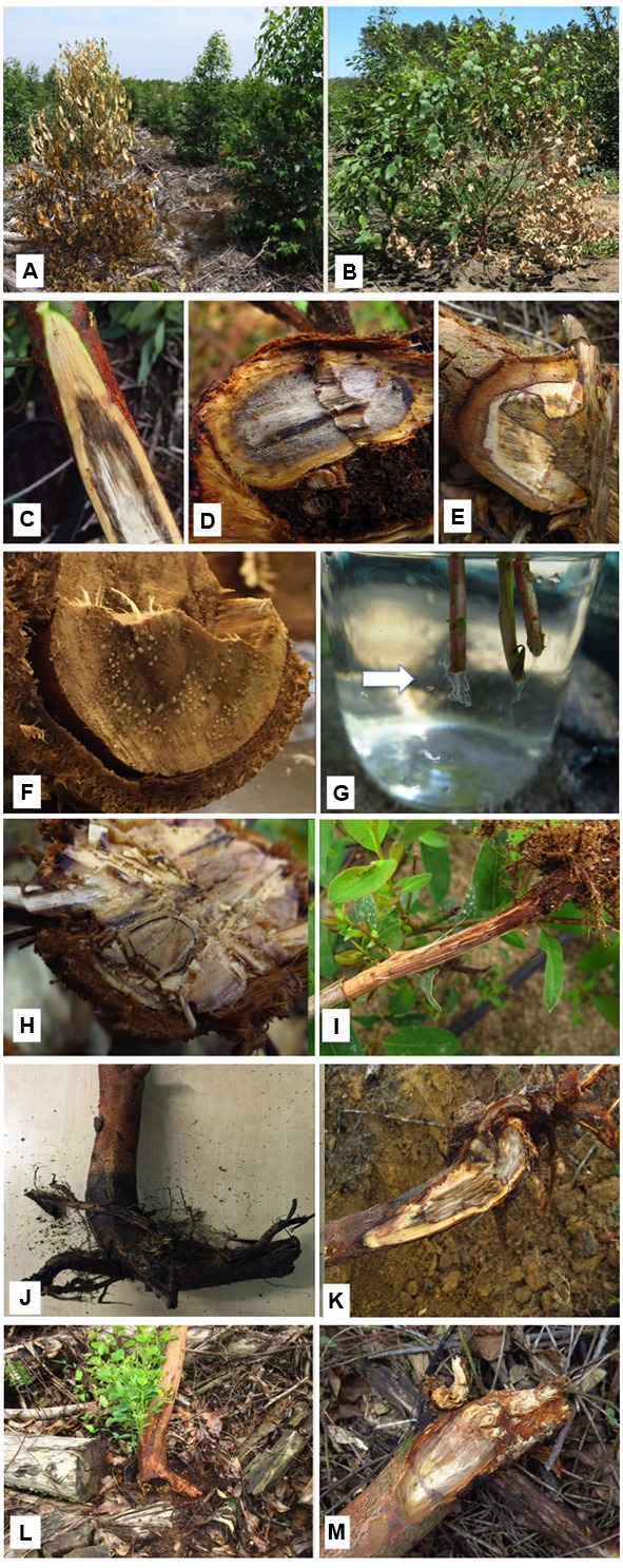
FIGURE 1.

Symptoms of bacterial wilt on eucalypts. (A) Entire tree showing signs of having died rapidly with leaves retained. (B) Infected tree commonly display partial death with one or a few branches wilted. This is linked to occlusion of parts but not all of the vascular tissue. (C) Internal discolouration of the vascular tissue of an infected tree. (D) Internal discolouration of the vascular tissue in a severely infected tree showing (exudation of bacteria. (E) Section of the base of a partially infected tree showing wood discoloration associated with an impacted root. (F) Bacterial exudateevident on the cut surface of an infected tree. (G) Bacteria seen exuding from the freshly cut stems (arrow) of infected nursery plants placed in water. (H) Section through the base of an asymptomatic tree but where smallareas of bacterial exudate are present. These trees with effective root systems typically recover without symptom development. (I) Poorly developed root system of an infected tree which would not be able to sustain itsgrowth. (J) J-rooting arising from poor planting practice typically associated with bacterial infection. (K) Section through the base of a tree showing compacted roots and discoloration associated with bacterial infection. (L) Epicormic shoots commonly develop at the bases of trees withknotted roots and that are also infected with bacteria. Stress associated with these poor root systems allows bacteria to proliferate. (M) Base of a young tree infected with bacteria but also with the root rot pathogen, Ganoderma philippii. Ralstonia pseudosolanacearum is commonly found in tree suffering fromthis root rot disease.)
