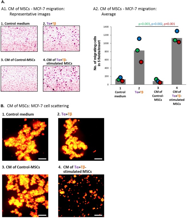
Figure 9.
Factors released by TNFα + TGFβ1-stimulated MSCs induce elevated migration and scattering of MCF-7 breast cancer cells. Human BM-derived MSCs were stimulated with vehicle (“Control-MSCs”) or TNFα (50 ng/ml) + TGFβ1 (10 ng/ml) [0.5% FBS-containing medium in panel (A) and FBS-free medium in panel (B)]; Tα + Tβ = TNFα + TGFβ1. In parallel, samples of “Control medium” (not exposed to MSCs), with or without the stimulating cytokines, were kept in identical conditions. Twenty-four hours later, all different media were filtered (0.45 µm pores) and applied to mCherry-expressing MCF-7 cells for 48 h (A) or 96 h (B). Then, functional assays were performed. CM = conditioned media. (A) Migration of MCF-7 cells toward 10% FBS-containing medium. (A1) Representative pictures of part of the high-resolution fields, ×40 magnification, of one of three independent experiments performed with MSCs of two different donors. (A2) Bar graph demonstrating the average number of cells migrating in each cell group, obtained in three independent experiments (total of five pictures/insert in each experiment); colored dots represent the number of migrating cells in each of these same three experiments, with corresponding color-coded p-values indicated. (B) Scattering of MCF-7 cells out of tumor spheroids. Scale bar = 200 µm. The pictures are representatives of n > 3 independent experiments, performed with MSCs of three different donors that have shown similar results.

