
Figure 3.
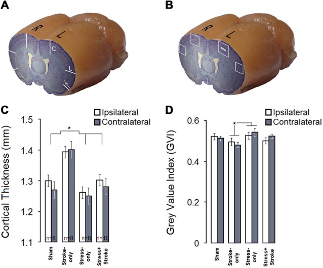
MPS and cortical ischemic lesion have differential effects on cortical thickness and neural density. (A) Coronal-sagittal view of a brain illustrating three cortical points (central, lateral, ventrolateral) used for cortical thickness measurements. (B) Stroke-only rats show greater cortical thickness compared to other experimental groups in both hemispheres. (C) Coronal-sagittal view of a rat brain illustrating bilateral regions of interest for quantitative cytoarchitectonics of absolute gray value index (GVI). White squares indicate four regions of interest (ROIs). (D) GVI measures indicated that MPS increased GVI in the contralateral hemisphere, whereas ischemic infarct resulted in reduced GVI in both hemispheres. Asterisks indicate significant differences: *p < 0.05.