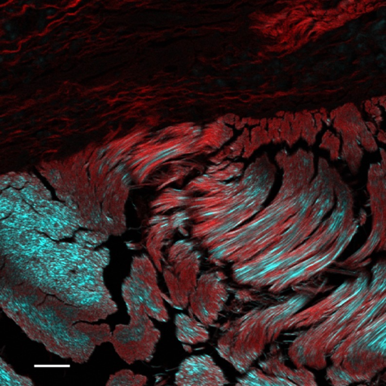
Fig. 1.
Kangaroo tail tendon allografted into mouse tissue. The sample has been stained with Sirius red, which gives the red signal by two-photon fluorescence. The type I collagen of the kangaroo tendon (bottom) shows a strong SH signal (cyan). Type III collagen from the mouse (above) stains strongly with the Sirius red but shows little SH signal at this setting. Scale bar 25 μm. Sample courtesy Allan Jones. See Cox et al. (2003)

