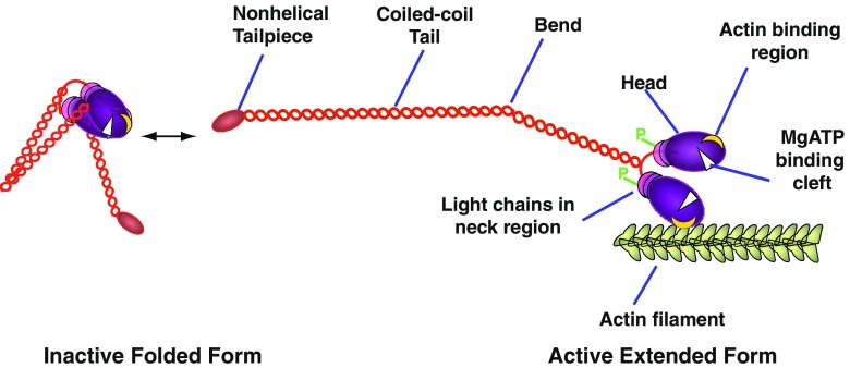
Fig. 1.
Schematic of Myosin 2 illustrating the phosphorylation-dependent reversible transition between the inactive folded monomer and the extended, assembly-competent, active form, the latter being able to interact with actin. Only one of two bends is shown in the extended myosin heavy chain. Phosphorylation sites (P) are located on each regulatory light chain in vertebrate myosin 2

