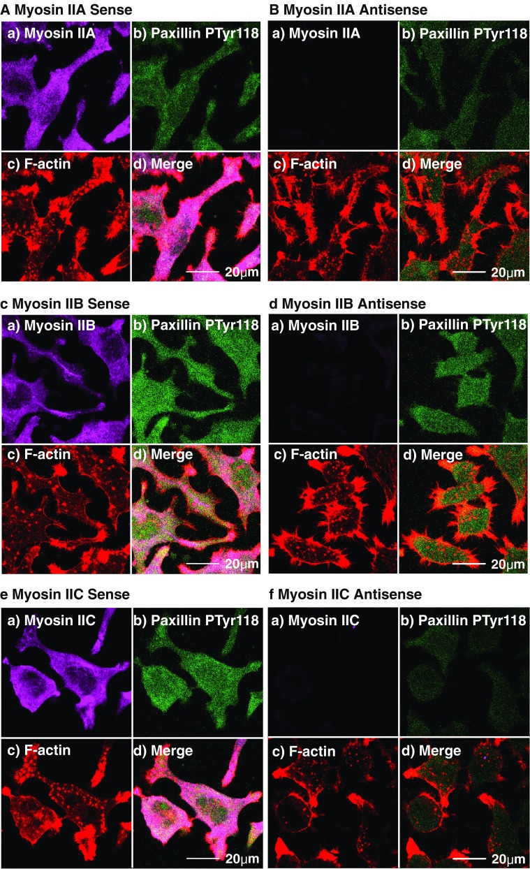
Fig. 4.

Targeted knockdown of M2A, M2B or M2C alters cell shape and/or cell adhesion. Paxillin phosphorylated on Tyr118 (an indicator of active adhesion) was observed relative to F-actin, M2A (a,b), M2B (c,d) or M2C (e,f) in Neuro-2A cells subsequent to 96-h exposure to either sense (a,c,e) or antisense (b,d,f) oligonucleotides targeting M2A (a,b), M2B (c,d) or M2C (e,f). All images obtained by CLS microscopy and are from confocal slices taken within 2 μm of the substratum to ensure inclusion of all structures associated with adhesion. Myosins pseudo-coloured in violet (Alexa-Fluor 633-labelled secondary) (a), phosphoTyr118-paxillin appears green (FITC-labelled secondary) (b), rhodamine-phalloidin F-actin is red (c) and merged images and scalebars are shown in (d). Note phenotypic changes as a result of M2B (d) and M2C (f) knockdowns, and corresponding attenuation of phosphoTyr118-paxillin expression with M2A (b) and M2C (f) knockdown
