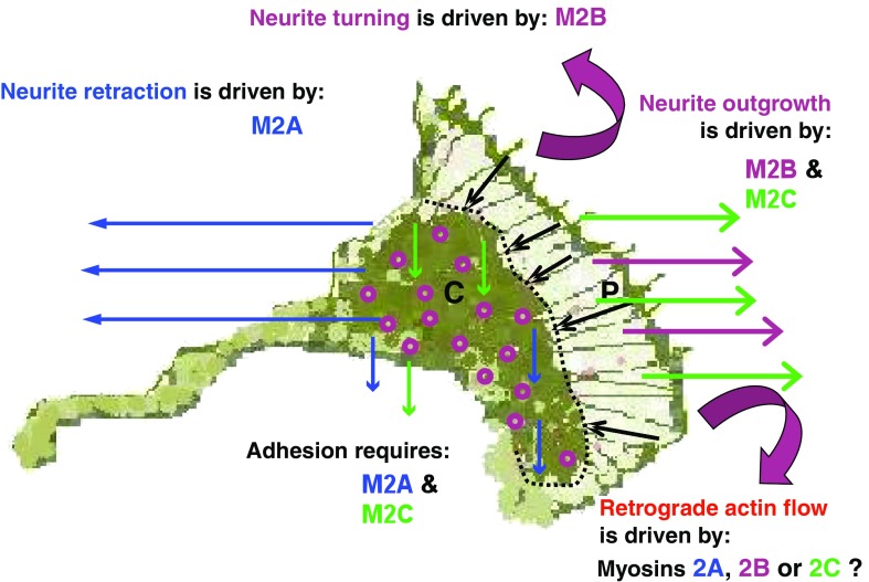
Fig. 5.
Schematic detailing the actions of M2A, M2B and M2C within a neuronal growth cone during development. M2A action is indicated by blue arrows, depicting M2A involvement in retraction and adhesion; M2B action is indicated by magenta arrows, depicting M2B involvement in outgrowth and turning; M2C action is indicated by green arrows, depicting M2C involvement in outgrowth and adhesion. Focal contacts are shown as purple “doughnuts”. P defines the peripheral zone of growth cone, separated from C, the central zone, by a dotted line. Black arrows indicate the direction of retrograde actin flow, known to be powered by one or more of the three myosin 2 motors

