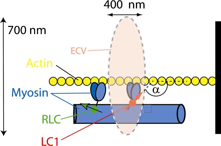
Fig. 1.
The principle of observing few molecules in muscle. ECV elliptical confocal volume. A small fraction of fluorescently labeled myosin is in ECV (here it is myosin that has been exchanged with LC1), which is characterized by a single transition dipole (red arrow). The dynamics of the fluorophore is determined by polarized fluorescence(polarized fluorescence) = [normalized difference between parallel (║) and perpendicular (┴) components the fluorescent light]. The probability distribution of polarized fluorescence is a measure of the extent of dispersion of orientations of lever arm orientations (Δα)

