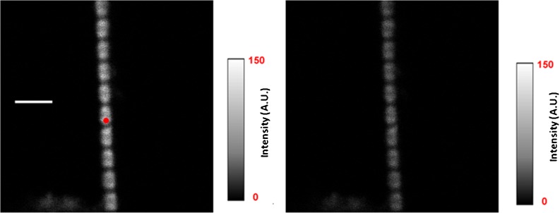
Fig. 2.
Intensity images of a rigor myofibril. The red circle is a projection of the confocal aperture on the sample plane (diameter 0.5 μm). Left image direction of polarization of exciting light is parallel (║I║, left image) and perpendicular (║I┴ , right image) to the axis of a myofibril. The intensity scales are in arbitrary units with 0 corresponding to black and 150 to white. Native myofibrillar LC1 was exchanged with 10 nM SeTau-LC1 . Scale bar 5 μm, sarcomere length 2.1 μm. Images were acquired on a PicoQuant Micro Time 200 confocal lifetime microscope. The sample was excited with a 635-nm pulsed laser and observed through a LP 650 filter

