Fig. 1.
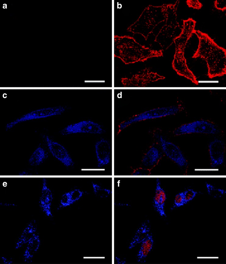
Confocal microscopy images of intracellular delivery of AuNCs. HeLa cells were treated with 11-mercaptoundecanonic acid-capped AuNCs (a, b) and peptide–functionalized AuNCs (c–f) for 1.5 h. The left panel shows fluorescence images of AuNCs only (blue), while the right panel shows colocalization images of AuNCs-treated cells that were counterstained with a membrance dye (wheat germ agglutinin-conjugated Alexa 594; b, d, red) and a nuclear dye (SYTO 59; f, red). Scale bar 25 μm. Reprinted with permission from the Royal Society of Chemistry (Lin et al. 2008)

