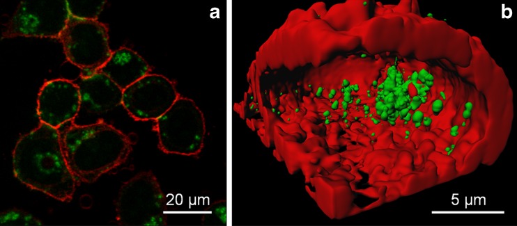
Fig. 2.
a Fluorescence images of HeLa cells after incubation with D-penicillamine-coated AuNCs (100 μg/ml) for 2 h, taken with 810-nm two-photon excitation. b Cross-section of a 3D image reconstruction, showing internalized AuNCs. AuNC fluorescence was recorded in a spectral window from 557 to 607 nm (green channel), whereas the emission of the membrane stain DiD was detected in a band from 647 to 703 nm (red channel)

