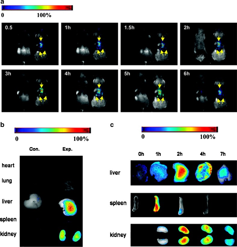
Fig. 5.
In vivo and ex vivo imaging female nude mice injected with ferritin-encapsulated AuNCs. a Whole body dorsal fluorescence images at different time points after Au NC injection. For each panel, the Au NC-injected mouse is shown on the right; a saline-injected control is shown on the left. b Fluorescence images of mouse organs 6 h after AuNCs injection. Control mice are shown on the left. c Fluorescence images of mouse organs at different time points after injection. The final concentration of AuNCs was 0.8 nmol/g body weight. The excitation filter transmitted from 576 to 621 nm; the emission was collected through a 635-nm long-pass filter. Reprinted with permission from American Chemical Society (Sun et al. 2011)

