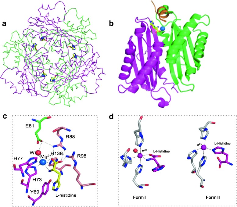
Fig. 4.
Structure of HutP-L-His-Mg2+ (PDB code: 1WPV). L-His and Mg2+ are shown as a yellow-colored stick model and a blue sphere model, respectively. Different HutP monomers are shown in different colors. a Cα backbone chain presentation of the HutP hexamer, L-His, and Mg2+, viewed along the threefold symmetry axis, b ribbon presentation of the HutP dimer, c L-His-Mg2+ binding site in the HutP-L-His-Mg2+ complex. Dotted lines Coordination interactions and hydrogen bonds, red sphere water molecule. d Alternative-binding modes of L-His with respect to the metal ions

