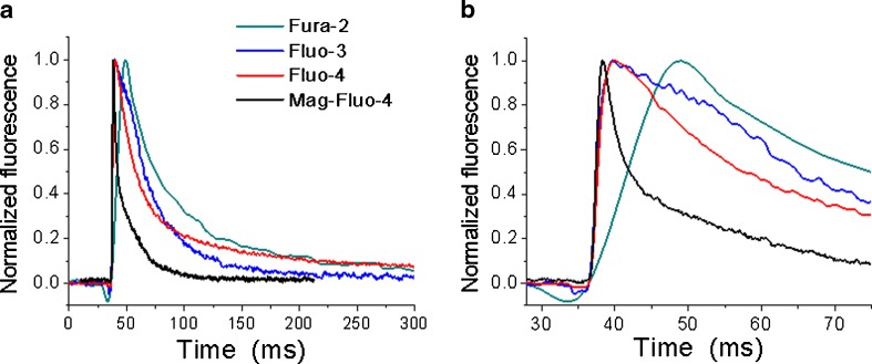
Fig. 1.
Comparison of single Ca2+ transients’ kinetics recorded in muscle fibres obtained by enzymatic dissociation of flexor digitorum brevis muscles from adult mice. Different cells were loaded with each of the Ca2+ dyes indicated in the figure and electrically stimulated. Ca2+ transients were recorded in an inverted fluorescence microscope using the appropriate set of filters, a photomultiplier and a Nikon amplifier. In (a), clear kinetic differences can be recognized, mostly derived from the different dissociation constants of the dyes used, being the fastest signal that obtained with Mag-Fluo-4 (black trace) and the slowest one that obtained with Fura-2 (green trace). In (b), the records are shown in an expanded time scale to better illustrate differences in the rising part of the signal

