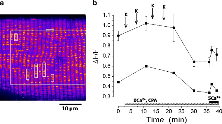
Fig. 5.
Increase in intramitochondrial Ca2+ during a sarcoplasmic reticulum (SR) depletion protocol in a flexor digitorum brevis fibre imaged in a confocal microscope. a A pseudocolor image of mitochondria loaded with Rhod-2. b Summarizes the time course of the mitochondrial mean Rhod-2 fluorescence variation in the regions of interest (white squares) marked in (a). The black circles represent the mean fluorescence in the small white squares in (a), and the black squares the mean fluorescence in the big white square in (a). The mitochondrial fluorescence increases during the protocol in which the cell is in absence of external Ca2+ and the SR Ca2+ ATPase is blocked by cyclopiazonic acid (CPA). When 5 mM Ca2+ external solution reaches the cell the mitochondria uptake part of the Ca2+ entering the fibre via the store operated Ca2+ entry mechanism. K indicates the moments in which an external solution with high K+ concentration was applied to elicit SR Ca2+ release in order to deplete this Ca2+ store

