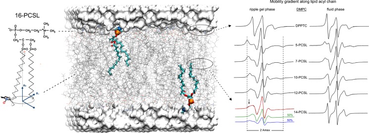
Fig. 1.
Spin labeling ESR from the membrane perspective and structural dynamics-line-shape correlations. a Dimiristoylphosphatidylcholine (DMPC) lipid bilayer (stick representation in gray) usually doped with 0.5–1.0 mol% of spin-labeled lipids containing a nitroxide radical attached to different positions along the lipid acyl chain, such as the spin label 16-PCSL on the left. A particular phospholipid is highlighted in licorice representation with the phosphorus and nitrogen atoms of the lipid head group colored in orange and blue, respectively. Different n-PCSL (n = 5, 7, 10, 12, 14, and 16) spin labels report on specific regions of the lipid bilayer. On the right, typical n-PCSL ESR spectra obtained for DMPC in the ripple gel phase (20 °C) and in fluid phase (35 °C). It is worth noting the lineshape changes of the spectra due to the mobility gradient experienced by the spin labels from the head group region down to the hydrophobic core of the lipid bilayer and also the lipid phase-dependence of the lineshape. Coexistence of two spin populations presenting different ordering and dynamics can also be detected by ESR, as shown by the 14-PCSL in the DMPC ripple gel phase. The lipid bilayer was built with CHARMM-GUI Membrane Builder (http://www.charmm-gui.org/input/membrane) (Jo et al. 2008; Wu et al. 2014) and rendered with Visual Molecular Dynamics (Humphrey et al. 1996). Adapted from (Basso et al. 2011) with permission

